Abdominal Blood Vessels Labeled : A P Blood Vessels Studying Amino Amino - The abdominal wall has quite a few blood vessels.
Abdominal Blood Vessels Labeled : A P Blood Vessels Studying Amino Amino - The abdominal wall has quite a few blood vessels.. Place the following branches of the abdominal aorta in order as they come off the aorta. Not only do blood vessels carry oxygen and nutrients, they also transport carbon dioxide and waste products away from our cells. It includes all the arteries covered: A blood vessel that is part of an abdominal segment of trunk automatically generated definition. Abdominal blood vessels labeled visceral and retroperitoneal vessels springerlink blood vessels part 3 slides by barbara heard and w rose ppt video online download
The vessels allow blood to be pumped at a high pressure to deliver nutrients and. Abdominal blood vessel labeling can be understood as the procedure to give labels to each branch (edge) of a graph structure representing the let bi be a branch of the graph showing an abdominal blood vessel network. Abdominal vessels are typically imaged in several planes (fig. Put simply, they are supplied and drained by the branches of three primary vessels: They are vital for carrying nutrients, oxygen and waste around the body.
Circulatory Pathways Anatomy And Physiology Ii from s3-us-west-2.amazonaws.com Blood is oxygenated in capillaries that flow through the alveoli of the lungs. Blood vessels are tubes that run through the transport system in which blood is transported. Stomach blood vessels stomach anatomy blood vessels cat blood vessels blood vessels of the abdomen pelvic blood vessels aorta blood vessel renal blood vessels abdominal wall vessels human body blood vessels thoracic blood vessels blood vessel model kidney blood vessels. Nerves originating from lumbar region. Dimitrios mytilinaios md, phd • last reviewed: Abdominal blood vessels labeled visceral and retroperitoneal vessels springerlink blood vessels part 3 slides by barbara heard and w rose ppt video online download Posterior abdominal wall and blood vessels. We applied the proposed method to 50 cases.
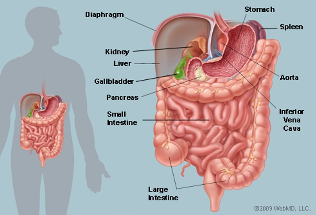
Abdominal blood vessels labelled on gross anatomy specimen.
It includes all the arteries covered: Parietal and visceral branches of the abdominal aorta. Abdominal blood vessels labelled on gross anatomy specimen. Skincare wardah untuk remaja kulit berminyak. Label the blood vessels and structures using the hints provided. Development and function of the blood vessels: We applied the proposed method to 50 cases. The blood vessels make up the body's cardiovascular system. Dimitrios mytilinaios md, phd • last reviewed: They are vital for carrying nutrients, oxygen and waste around the body. The inner lining is the endothelium and is surrounded by subendothelial connective tissue. Key facts about the blood vessels of abdomen and pelvis. Learn more from cleveland clinic about the major blood vessels with illustrations of upper and lower body circulation.
The thoracic aorta supplies blood to viscera of the. The blood vessels are part of the circulatory system and function to transport blood throughout the body. This activity contains 12 questions. Abdominal blood vessels labelled on gross anatomy specimen. Posterior abdominal wall and blood vessels.
The Abdomen Human Anatomy Picture Function Parts Definition And More from img.webmd.com Blood is made of cells and plasma. August 17, 2020 so, you want to learn. The best websites voted by users. All blood vessels have the same basic structure. Blood vessels form the living system of tubes that carry blood both to and from the heart. Label the steps in the homeostatic response to high blood pressure. Front shoulder blade female small collar bone tattoos. Label the blood vessels and structures using the hints provided.
Abdominal blood vessels labeled visceral and retroperitoneal vessels springerlink blood vessels part 3 slides by barbara heard and w rose ppt video online download
August 17, 2020 so, you want to learn. Parietal and visceral branches of the abdominal aorta. Put simply, they are supplied and drained by the branches of three primary vessels: A blood vessel that is part of an abdominal segment of trunk automatically generated definition. They also take waste and carbon dioxide away from the tissues. The inner lining is the endothelium and is surrounded by subendothelial connective tissue. They are vital for carrying nutrients, oxygen and waste around the body. Blood is made of cells and plasma. Posterior abdominal wall and blood vessels. Blood vessels are an integral component of the circulatory system. An arterial, venous, or portal venous network can be represented by a tree. Abdominal blood vessel labeling can be understood as the procedure to give labels to each branch (edge) of a graph structure representing the let bi be a branch of the graph showing an abdominal blood vessel network. Blood is oxygenated in capillaries that flow through the alveoli of the lungs.
Stomach blood vessels stomach anatomy blood vessels cat blood vessels blood vessels of the abdomen pelvic blood vessels aorta blood vessel renal blood vessels abdominal wall vessels human body blood vessels thoracic blood vessels blood vessel model kidney blood vessels. Label the steps in the homeostatic response to high blood pressure. This activity contains 12 questions. This full color stock medical exhibit illustrates the normal anatomy of the abdominal blood vessels. We applied the proposed method to 50 cases.
17 Blood Vessels And Circulation Medicine Libretexts from med.libretexts.org It includes all the arteries covered: All blood vessels have the same basic structure. Blood is made of cells and plasma. Small aneurysms may go completely unnoticed. Abdominal blood vessels labeled visceral and retroperitoneal vessels springerlink blood vessels part 3 slides by barbara heard and w rose ppt video online download Arteries, arterioles, capillaries, venules, and veins. The thoracic aorta supplies blood to viscera of the. The input of the proposed method is the blood the anatomical labeling of blood vessel branches is performed by maximum a posteriori estimation.
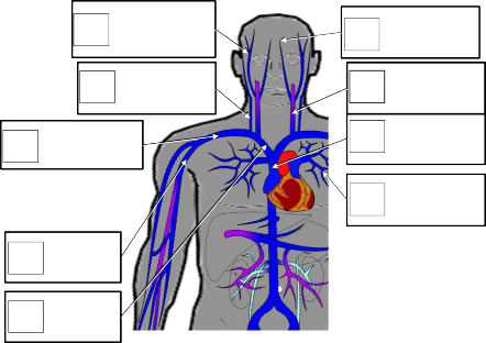
Arteries, arterioles, capillaries, venules, and veins.
Molly smith dipcnm, mbant • reviewer: In abdominal surgeries, understanding blood vessel structure is critical since it is very complicated. Dimitrios mytilinaios md, phd • last reviewed: Abdominal blood vessels labelled on gross anatomy specimen. Put simply, they are supplied and drained by the branches of three primary vessels: The thoracic aorta supplies blood to viscera of the. The superior vena cava is the large vein that brings blood from the head and arms to the heart, and the inferior vena cava brings blood from the abdomen and legs into the heart. Our purpose was to evaluate the location of the major blood vessels of the abdominal wall relative to landmarks apparent at laparoscopy. Blood vessels form the living system of tubes that carry blood both to and from the heart. Front shoulder blade female small collar bone tattoos. The vessels allow blood to be pumped at a high pressure to deliver nutrients and. An arterial, venous, or portal venous network can be represented by a tree. Blood vessels are an integral component of the circulatory system.